Animal Cell Under An Electron Microscope : Electron Microscopic Study Of Cell And Organelles Important / The largest known animal cell is the ostrich egg, which can stretch over 5.1 inches across and weighs about 1.4 kilograms.
byEna Jinenez-
0
Animal Cell Under An Electron Microscope : Electron Microscopic Study Of Cell And Organelles Important / The largest known animal cell is the ostrich egg, which can stretch over 5.1 inches across and weighs about 1.4 kilograms.. Observation of euglena under more powerful electron microscopes have revealed the presence of an ornamented pellicle under the plasma membrane. It is also freely scattered throughout the entire cytoplasm and it will depend on the cell whether it is a plant or animal or bacterial cell. In order to view the organelles an electron microscope is needed. Moreover, because of their flexible nature, they also facilitate movement. Animal cells range in size from a few microscopic microns to few millimetres.
The largest known animal cell is the ostrich egg, which can stretch over 5.1 inches across and weighs about 1.4 kilograms. They use electrons (negatively charged electrical particles) to magnify objects up to two million times. Sems do not use light waves; Animal cells range in size from a few microscopic microns to few millimetres. The presence of this thin protein layer protects the their cell membrane and also helps in maintaining their shape.
Illustrate Only A Plant Cell As Seen Under Electron Microscope How Its Different From Animal Cell Biology Q A Doubtnut from doubtnut-static.s.llnwi.net Histology is the microscopic counterpart to gross anatomy, which looks at larger structures visible without a microscope. Preparing a wet mount of a specimen is the technique typically used to view plant and animal cells using a microscope.this page provides step by step instructions on slide preparation as well as videos at the bottom of page. In order to view the organelles an electron microscope is needed. Sems do not use light waves; Observation of euglena under more powerful electron microscopes have revealed the presence of an ornamented pellicle under the plasma membrane. Moreover, because of their flexible nature, they also facilitate movement. Below is a picture of two euglena organisms, seen just after reproduction. Animal cells range in size from a few microscopic microns to few millimetres.
The presence of this thin protein layer protects the their cell membrane and also helps in maintaining their shape.
With modern microscopes, the processes behind cell theory can easily be viewed and studied. Moreover, because of their flexible nature, they also facilitate movement. The largest known animal cell is the ostrich egg, which can stretch over 5.1 inches across and weighs about 1.4 kilograms. Below is a picture of two euglena organisms, seen just after reproduction. Histology is the microscopic counterpart to gross anatomy, which looks at larger structures visible without a microscope. Mostly, the ribosomes are found to be bound to the endoplasmic reticulum and the nuclear envelope. Since this microscope produces a visible, clear image of small organelles, in an electron microscope there is no need for reagents to see the virus or harmful cells, resulting in a more efficient way to detect pathogens. The bulb of an onion is formed from modified leaves. Histology, also known as microscopic anatomy or microanatomy, is the branch of biology which studies the microscopic anatomy of biological tissues. One of the latest discoveries made about using an electron microscope is the ability to identify a virus. Onion cells under a microscope requirements, preparation and observation. The presence of this thin protein layer protects the their cell membrane and also helps in maintaining their shape. Observation of euglena under more powerful electron microscopes have revealed the presence of an ornamented pellicle under the plasma membrane.
The presence of this thin protein layer protects the their cell membrane and also helps in maintaining their shape. Mostly, the ribosomes are found to be bound to the endoplasmic reticulum and the nuclear envelope. While photosynthesis takes place in the leaves of an onion containing chloroplast, the little glucose that is produced from this process is converted in to starch (starch granules) and stored in the bulb. Since this microscope produces a visible, clear image of small organelles, in an electron microscope there is no need for reagents to see the virus or harmful cells, resulting in a more efficient way to detect pathogens. Preparing a wet mount of a specimen is the technique typically used to view plant and animal cells using a microscope.this page provides step by step instructions on slide preparation as well as videos at the bottom of page.
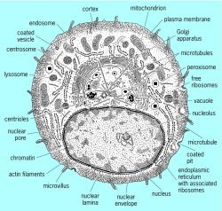
Animal Cell Microscope Labeled Animal Cell Diagram from niu.quibicatnap.fun The largest known animal cell is the ostrich egg, which can stretch over 5.1 inches across and weighs about 1.4 kilograms. Below is a picture of two euglena organisms, seen just after reproduction. Histology is the microscopic counterpart to gross anatomy, which looks at larger structures visible without a microscope. One of the latest discoveries made about using an electron microscope is the ability to identify a virus. Mostly, the ribosomes are found to be bound to the endoplasmic reticulum and the nuclear envelope. Histology, also known as microscopic anatomy or microanatomy, is the branch of biology which studies the microscopic anatomy of biological tissues. A great example of watching cell theory in action can be accomplished by putting a drop of pond water under a microscope. Preparing a wet mount of a specimen is the technique typically used to view plant and animal cells using a microscope.this page provides step by step instructions on slide preparation as well as videos at the bottom of page.
Histology is the microscopic counterpart to gross anatomy, which looks at larger structures visible without a microscope.
One of the latest discoveries made about using an electron microscope is the ability to identify a virus. Sems do not use light waves; The largest known animal cell is the ostrich egg, which can stretch over 5.1 inches across and weighs about 1.4 kilograms. Preparing a wet mount of a specimen is the technique typically used to view plant and animal cells using a microscope.this page provides step by step instructions on slide preparation as well as videos at the bottom of page. Mostly, the ribosomes are found to be bound to the endoplasmic reticulum and the nuclear envelope. Histology, also known as microscopic anatomy or microanatomy, is the branch of biology which studies the microscopic anatomy of biological tissues. While photosynthesis takes place in the leaves of an onion containing chloroplast, the little glucose that is produced from this process is converted in to starch (starch granules) and stored in the bulb. With modern microscopes, the processes behind cell theory can easily be viewed and studied. Moreover, because of their flexible nature, they also facilitate movement. It is also freely scattered throughout the entire cytoplasm and it will depend on the cell whether it is a plant or animal or bacterial cell. In order to view the organelles an electron microscope is needed. Histology is the microscopic counterpart to gross anatomy, which looks at larger structures visible without a microscope. They use electrons (negatively charged electrical particles) to magnify objects up to two million times.
The presence of this thin protein layer protects the their cell membrane and also helps in maintaining their shape. Animal cells range in size from a few microscopic microns to few millimetres. A great example of watching cell theory in action can be accomplished by putting a drop of pond water under a microscope. Below is a picture of two euglena organisms, seen just after reproduction. One of the latest discoveries made about using an electron microscope is the ability to identify a virus.
Cell Structure Article About Cell Structure By The Free Dictionary from img.tfd.com Sems do not use light waves; Onion cells under a microscope requirements, preparation and observation. Moreover, because of their flexible nature, they also facilitate movement. The bulb of an onion is formed from modified leaves. Since this microscope produces a visible, clear image of small organelles, in an electron microscope there is no need for reagents to see the virus or harmful cells, resulting in a more efficient way to detect pathogens. While photosynthesis takes place in the leaves of an onion containing chloroplast, the little glucose that is produced from this process is converted in to starch (starch granules) and stored in the bulb. The largest known animal cell is the ostrich egg, which can stretch over 5.1 inches across and weighs about 1.4 kilograms. Below is a picture of two euglena organisms, seen just after reproduction.
Histology is the microscopic counterpart to gross anatomy, which looks at larger structures visible without a microscope.
The bulb of an onion is formed from modified leaves. The presence of this thin protein layer protects the their cell membrane and also helps in maintaining their shape. Sems do not use light waves; Histology is the microscopic counterpart to gross anatomy, which looks at larger structures visible without a microscope. They use electrons (negatively charged electrical particles) to magnify objects up to two million times. Below is a picture of two euglena organisms, seen just after reproduction. Onion cells under a microscope requirements, preparation and observation. It is also freely scattered throughout the entire cytoplasm and it will depend on the cell whether it is a plant or animal or bacterial cell. A great example of watching cell theory in action can be accomplished by putting a drop of pond water under a microscope. Mostly, the ribosomes are found to be bound to the endoplasmic reticulum and the nuclear envelope. The largest known animal cell is the ostrich egg, which can stretch over 5.1 inches across and weighs about 1.4 kilograms. With modern microscopes, the processes behind cell theory can easily be viewed and studied. Preparing a wet mount of a specimen is the technique typically used to view plant and animal cells using a microscope.this page provides step by step instructions on slide preparation as well as videos at the bottom of page.